What are characteristics of a living cell?
Growth and reproduction are the characteristics of cells.
Have you ever wondered how you started your life as a single cell and grew up to be an individual made up of millions of cells?
Well, this was possible because cells are capable of dividing and multiplying. Rudolf Virchow had added to the cell theory ‘Omnis cellula-e-cellula’ which means that all cells arise from pre-existing cells.
Do you know how new cells arise from old ones? It is by the process of cell division. Most cell divisions in our body result in the formation of daughter cells which are exact replicas of the parent cells, except for the cell division which result in the formation of gametes. In such a division, the daughter cells receive only half the amount of genetic material from the parent cell.
Why do cells need to divide?
Once a cell grows to a particular size, it divides to avoid getting bigger. Transport of substances in and out of the cell becomes inefficient as the cell grows larger. Thus, to keep a small size, cells divide after a point of time.
Also, the DNA content of the cell will remain the same, even if it continues to grow larger. But with larger size, it becomes difficult for the DNA of the cell to keep up with the demands of a larger cell. Thus, the cell divides to keep the size small enough for the cellular DNA to be able to meet all its demands.
Cell Cycle and Cell Division
Cell division is the process by which a parent cell divides into two or more daughter cells.
Cell division is essential for growth, repair and reproduction. Many cell cycles transform a single cell to a multicellular organism.
Cell division is an important life process that helps us to grow. But what happens once we grow up? Do our cells stop dividing? Well no, they don’t.
Cell division not only helps in growth but also helps in repair and regeneration of old and worn out cells in the body.
But do all our body cells require repair and regeneration at the same rate? No, they don’t. While the cells of your skin divide once in every month, the ones in your liver cells divide only once a year. How is this controlled?
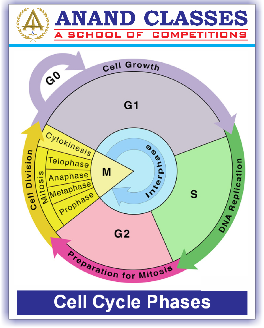
In fact, the entire process of cell division is a complicated event which involves growth of the cell, replication of cell DNA and the actual division of the cell, all sequences are coordinated in a controlled manner. This entire sequence of events is known as the cell cycle.
What is Cell Cycle ?
The sequence of events takes place in a cell by which a cell duplicates its genome (DNA), synthesizes the cell’s other constituents such as cytoplasm and organelles & their distribution and subsequently divides into two daughter cells is called the cycle of cells.
 The cell cycle was discovered by Prevost and Dumas (1824) while studying the cleavage of zygote of Frog. It is a series of stages a cell passes through, to divide and produce new cells.
- The cell cycle comprises three coordinated processes of cell division, DNA replication, and cell growth.
- Events happening in a cell cycle is genetically controlled
Cell cycle period can vary from organism to organism, and from cell type to cell type. (E.g., 90 minutes in the yeast cell cycle, 24 hours in humans.)
- The cell cycle is divided into two major phases : Interphase and M phase
The cell cycle has multiple stages and multiple checkpoints to regulate the transition from one stage to the next.
These checkpoints are genetically controlled by the formation of certain proteins which decide when the cell should start dividing and when it should stop.
Duration of cell cycle
The period taken to complete one cell cycle, that is, from the beginning of one cell division to the beginning of the next, is known as the generation time. Duration of cell cycle varies from cell to cell and from organism to organism. A human cell can complete a cell cycle in 24 hours while a yeast cell takes 90 minutes.
Phases of cell cycle
Cell cycle occurs in two phase :
- Interphase : A phase of cell cycle when the cell prepares itself for cell division.
- M phase (Mitosis) : A phase of cell cycle when the actual division occurs.
Interphase
Before the cell divides, it must have all the necessary requirements for producing two daughter cells. Production of necessary requirements of the cell cycle occurs at the interphase stage.
Interphase is the time lapse between two successive M phases of cell division. In this phase the cell prepares for division, grows and DNA replication takes place.
The interphase is a phase of non-apparent division of the cell. It is also called as Resting phase because in this phase cell is only prepare for division not divide.
Interphase is divided into 3 additional phases: the “G1” phase, the “S” phase, and the “G2” phase.
Interphase stage occupies 95% of the total duration of the cell cycle. If a cell takes 24 hours to complete a cell cycle, the interphase stage takes 23 hours of the total 24 hours.
Important Interphase Stage Tasks (Siginificance)
There are a few important tasks that a cell needs to accomplish at interphase stage :
- Growth in the size of cells in terms of organelles and membrane such that it can distribute enough cellular constituents to the two daughter cells.
- Creating a copy of the genetic material, i.e, DNA so that genetic material can be distributed among the daughter cells.
- Synthesis and storage of ATP to be used as an energy currency for the various stages of cell division.
- Division of the centrosome and production of two centriole pairs in case of animal cells.
Stages of interphase
Interphase is divided into 3 stages:
- G1 Phase (Gap 1)
- S Phase (Synthesis phase)
- G2 Phase (Gap 2)
 G1 Phase (Gap 1)
The phase between mitosis and S phase of the cell cycle. The cell is metabolically active in this phase and grows larger physically without replicating its DNA.
Production of nutrients and protein required for S phase are synthesised in this phase. Copies of cell organelles are also produced.
The duration of this phase can vary from a few minutes to several days. Cells which divide very rarely have a longer G1 phase compared to the ones which divide very frequently.
S Phase (Synthesis Phase)
The phase between G1 and G2 phase of the cell cycle. The S phase or synthesis phase involves replication of DNA and production of a copy of the genetic material of the cell.
DNA is present in the form of condensed chromosomes in the nucleus of the cell. During S Phase, If the initial quantity of DNA in the cell is denoted as 2N, then after replication it becomes 4N by end of synthesis phase. However the number of chromosomes does not vary, viz., if the number of chromosomes during G1 phase was 2n, it will remain 2n at the end of S phase. The centriole also divides into two centriole pairs in the cells which contain centriole.
hence, in this phase, the DNA content of the cell doubles and centriole duplicates. It is important to note that the chromosome number remains the same.
Along with DNA, synthesis of histone proteins, assembly of kinetochore units and formation of nucleosomes also occurs at this stage. S phase commits the cell to divide.
DNA is known as the blueprint of life as it holds all the information required to build the cell and run all the activities of the cell. Daughter cells inherit the genetic material from the parent cell and in order to distribute genetic material equally among daughter cells, a copy of existing genetic material is required.
G2 Phase (Gap 2)
Starts after the S phase of the cell cycle. During this phase the cell size increases, nucleus grows in size, and ATP production and storage continues. There is intensive synthesis of RNA and proteins, Division and multiplication of cell organelles like mitochondria, chloroplasts, etc occurs during the G2 phase.
During this phase, the RNA, proteins, other macromolecules required for multiplication of cell organelles, spindle formation, and cell growth are produced as the cell prepares to go into the mitotic phase.
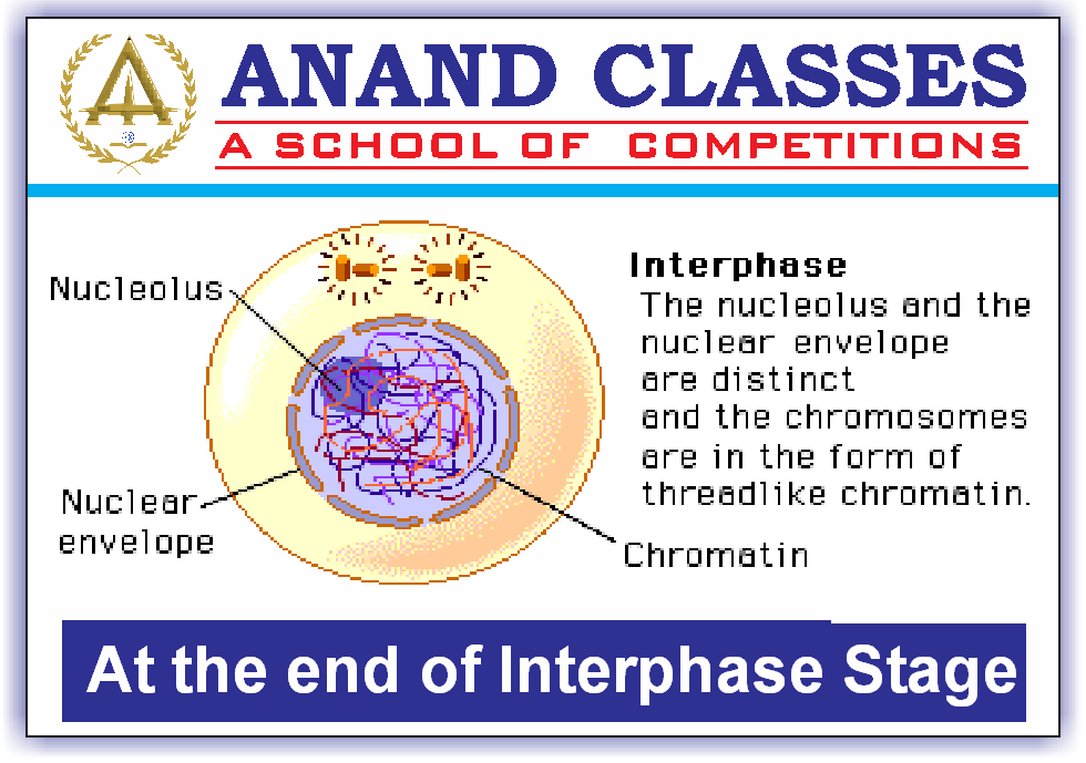
At the end of Interphase, a cell that is ready to divide looks like this:
 G0 Phase/Gap Phase/Quiescent stage
In an adult human being, Not all cells in our body divide and there are also many cells which divide occasionally (e.g. heart cells divide occasionally only to replace injured and dead cells). Cells which do not divide further enter into the inactive stage called G0 phase after the G1 phase of the cell cycle.
Cells in this phase, although metabolically active, do not divide unless and until are directed to do so as per the requirement of the cell.
Cells such as cardiac cells, nerve cells, skeletal muscles, RBCs do not divide after their growth and differentiation and remain in the G0 phase forever.
M Phase
The most dramatic period of the cell cycle. It is the phase of the cell cycle that occurs after the interphase. This is the mitotic phase as the cell undergoes a complete reorganization to give birth to a progeny that has the same number of chromosomes as the parent cell.
In this phase actual cell division occurs. Nuclear division happens in this phase, where daughter chromosomes are separated. The number of chromosomes in the parent and daughter cells remains the same so it is also known as equational division.
It involves the mitotic or meiotic division of a cell. Mitotic division equally distributes the chromosomes in the two daughter cells whereas meiotic division distributes only half the number of chromosomes into the daughter cells.
Important points regarding Mitosis :
- Mitosis mostly occurs in the diploid somatic cells of animals with few exceptions, haploid male drone of honey bees
- In plants, mitosis happens in both haploid and diploid cells
- Mitosis is responsible for genetic continuity and growth and repair of multicellular organisms
- In humans, the epithelial lining, lining of gut and blood cells are replaced continuously
- In plants, meristematic tissues divide continuously throughout their life
- Mitosis accounts for the asexual reproduction or vegetative propagation, where identical individuals are formed
Stages of mitosis
Mitosis is divided into two stages:
- Karyokinesis
- Cytokinesis
Karyokinesis
The word karyokinesis is derived from two words:
Karyon – Means nucleus
Kinesis – Means movement
This stage of mitosis involves division of nucleus which occurs through following stages:
- Prophase
- Metaphase
- Anaphase
- Telophase
Cytokinesis
The word Cytokinesis is derived from two words:
Cytos – Means cell
Kinesis – Means movement
Karyokinesis, i.e. nuclear division is followed by cytokinesis, i.e. division of the cytoplasm to give rise to two daughter cells.
This stage of mitosis involves division of cytoplasm. After the segregation of chromosomal material, the next step is the division of cytoplasmic material of the cell. The process of cytokinesis differs in a plant cell and animal cell. In animal cells it occurs by furrowing of the cell membrane whereas in plant cells it occurs by cell plate formation.
Karyokinesis stage of the mitotic phase occurs in four sequential stages:
- Prophase
- Metaphase
- Anaphase
- Telophase
Prophase (longest stage)
This first stage of karyokinesis of mitosis occurs after the S and G2 phases of interphase.
Prophase is marked by the beginning of condensation of chromosomal material. New DNA molecules formed are condensed with chromosomal material in this phase. Compact mitotic chromosomes are formed. Chromosomes are composed of two chromatids at the centromere, which undergoes duplication. The centrosome, which had undergone replication during S Phase, now begins to move towards the opposite pole of the cell.
 In this phase following events happen :
- Chromosomes untangle and condense
- Two chromatids attached to the centromere can be seen clearly
- Each of the duplicated centrosomes radiates microtubules (asters)
- Mitotic apparatus constitutes spindle fibres and asters
- Spindle fibres emerge from centrosomes
- Golgi bodies, nucleolus, endoplasmic reticulum and nuclear membrane disappear
Metaphase
In this phase, the centromere holds two sister chromatids. Kinetochores, small disc-shaped structures, are formed at the surface of the centromeres. They are attached to the chromosomes through the spindle fibres. Later, the chromosomes get aligned through spindle fibres to both poles of the spindle equator.

- Complete disintegration of the nuclear envelope
- Two sister chromatids attached by the centromere aligned at the equator, i.e. metaphase plate
- Each chromatid is attached to spindle fibres from opposite poles at kinetochores
- Chromosomes spilt up and arrange themselves on the equator to form metaphase plate (equatorial plate).
- Spindle fibres are microtubules. Chromosomal fibres, (discontinuous and run from pole to centromere) and supporting fibres, (continuous and run from pole to pole), arrange in a cell.
- The centromere lies at the equator with arms facing the poles.
This is the stage where chromosomal morphology is most easily studied.
Anaphase (smallest stage)
- Each chromosome assembled at the metaphase plate is split simultaneously at the beginning of anaphase, and the two daughter chromatids begin to migrate towards the two opposite poles.
- When each chromosome travels away from the equatorial plate, the middle of each chromosome is towards the pole and therefore at the front edge, with the chromosome’s arms trailing behind it.
- Sister chromatids now become the daughter chromosomes
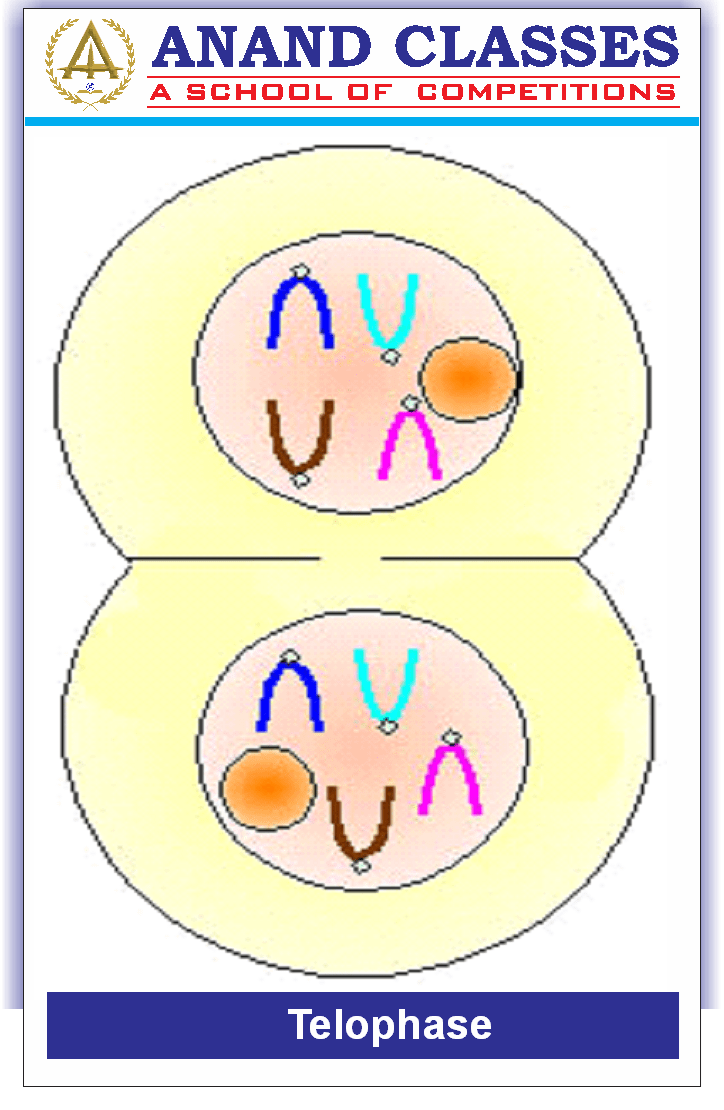
 Telophase (reverse of prophase)
- Chromosomes cluster at opposite poles and decondense
- Nuclear envelope develops around each cluster of chromosomes and two daughter nuclei are formed
- The nucleolus, endoplasmic reticulum and Golgi apparatus are reformed
 Cytokinesis
Cytokinesis is initiated in late the anaphase. It is different for plants and animals.
(i) Cytokinesis in animals
Following karyokinesis, a separate process called cytokinesis splits the cell itself into two daughter cells.
- Separation of cytoplasm takes place after two nuclei are formed. Cell organelles get distributed between daughter cells.
Cytokinesis in animal cells starts as a ring of actin filaments forms at the metaphase plate. The ring contracts which form a cleavage furrow inside the plasma membrane.
- The furrow slowly deepens, and eventually joins the middle separating the cytoplasm into two cells.
- The cytokinesis in animal cell occurs in the centripetal order.
(ii) Cytokinesis in plants
The cell plate formation takes place because the constriction or even furrow is not possible as the cell wall is rigid. Many Golgi vesicles and spindle microtubules arrange themselves on equator and the cell has a Phragmoplast. It may also have the deposits of fragments of ER. Golgi vesicles membranes fuse and form a plate like structure which is called as the cell plate. Golgi vesicles then secret pectates of calcium and magnesium. The cell plate modifies into the middle lamella.
Cellular cells of plants undergo cytokinesis via plate cells. Wall formation in the cell plate method starts at the center of the cell and extends towards the current lateral walls.
The formation of the new cell wall begins with the formation of a simple precursor called the cell plate representing the middle lamella between two adjacent cells ‘ walls..
Organelles such as mitochondria and plastids are distributed between the two daughter cells at the time of cytoplasmic division.
- The cytokinesis of plant cells occur in the centrifugal order (cell plate formation is from centre to periphery).
And after that, the eukaryotic cell cycle is initiated again staring with the G1 phase of interphase.
Note : In some organisms like fungi, algae and plant cells, cytokinesis is not immediately followed by karyokinesis and the multinucleate stage (disease) is formed known as a syncytium, e.g. liquid endosperm in coconut, coenocytic hyphae of Rhizopus, etc.
Significance of Mitosis
- Mitosis is the equational division is a common division method for the diploid cells only. However, some lower plants and social insects which have haploid cells, also use mitosis for division. The significance of this division is essential to understand in the life of an organism.
- Mitosis results in the production of diploid daughter cells which have identical genetic chromosome number. The multicellular organisms grow due to the mitosis.
- Cell growth often results in disturbing the usual ratio of the nucleus and the cytoplasm. Thus, the cell divides and restores the nucleo-cytoplasmic ratio.
- A very significant contribution is that a cell is repaired. Best examples are the cells of the upper epidermis layer, cells of the gut lining, and blood cells being replaced constantly.


 G1 Phase (Gap 1)
 G0 Phase/Gap Phase/Quiescent stage
 Cytokinesis